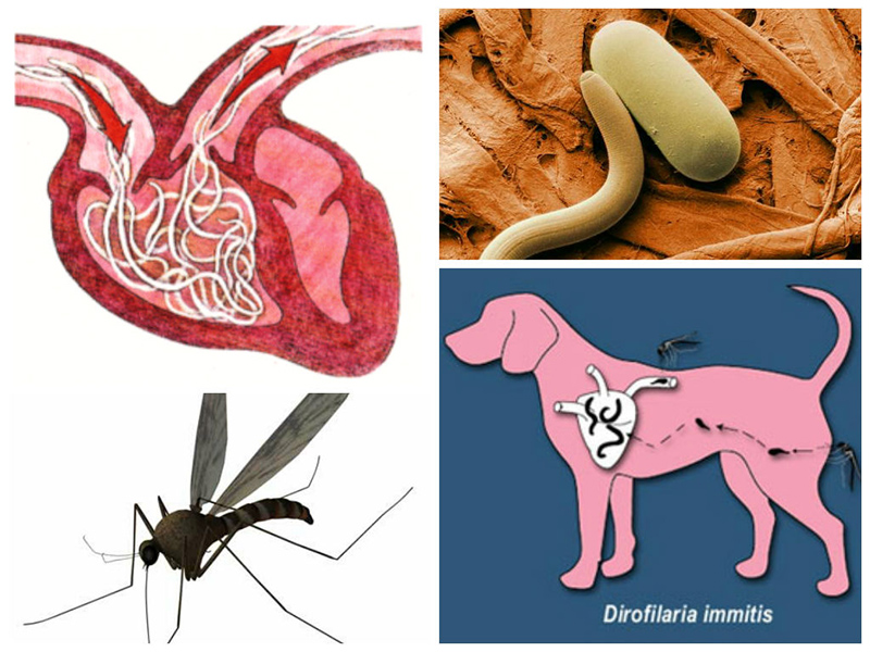
Dirofilariasis is a highly dangerous disease caused by roundworms from the nematode group, called dirofilaria. Adults are long (up to 40 cm), thin worms about 1.5 mm in diameter, and come in two varieties: immitis and repens. The former are more dangerous, as they infect the right ventricle of the heart and the pulmonary arteries, while the latter parasitize under the skin and inside the mucous membrane of the eye. Dirofilariasis affects not only dogs but also cats and humans. Mosquitoes (of the genus Aedes) are considered carriers of the disease, and the disease itself is often fatal.
Content
Causes of heartworm disease in dogs
A mosquito that bites an already infected dog becomes a temporary repository for parasite larvae (microfilariae), where they undergo several stages of development. Once the carrier insect bites a healthy animal, the individuals that have grown to the invasive stage enter the bloodstream, where they continue their further development. The maturation period of the parasite larvae inside the mosquito can range from 10 days to 1 month (depending on the ambient temperature).
Dirofilariasis (from the Latin "diro, filum" - "evil thread") is a helminthic disease caused by nematodes of the genus Dirofilaria.
In addition to dogs, other species can also be susceptible to infection: wolves, foxes, coyotes, domestic and wild cats, ferrets, muskrats, sea lions, coatis
Once in a dog's body, heartworms continue to grow for 5-7 months, after which they begin to actively reproduce. Adult heartworms of the immitis species, after completing their final stage of development, migrate throughout the bloodstream and accumulate in the heart or pulmonary artery. Heartworms repens do not enter the bloodstream and remain under the animal's skin, continuing to reproduce. One adult heartworm lives in a dog's body for approximately 5-7 years, and there may be up to 250 of them.
Parasites can, in isolated cases, be found in the abdominal cavity, brain, and muscle tissue.
Symptoms
Cardiac and subcutaneous forms of dirofilariasis have different symptoms and progression. The initial stage of the disease is often asymptomatic. The pet simply becomes slightly less active, its appetite decreases, and in the case of a subcutaneous infection, a noticeable raised lump appears on the surface of the skin.
Signs of cardiopulmonary damage:
- loss of appetite;
- elevated temperature;
- shortness of breath, difficulty breathing;
- weakness, apathy;
- severe swelling of the paws;
- expectoration of bloody sputum;
- fatigue and lethargy;
- slight cough.
In the Republic of Bashkortostan, infection rates among service dogs reach 25–30%, and when they are kept in enclosures in a limited area and without preventive measures, the rate rises to 90%.
Sometimes, an animal is diagnosed with kidney failure, liver damage, and ascites simultaneously. Associated symptoms of advanced disease include cyanosis of the mucous membranes, loss of consciousness, and wheezing. Heart damage is the most life-threatening condition for a dog. While anthelmintic medications may be effective in the initial stages, in the later stages, taking medications can actually cause harm. After all, dead parasites are unable to leave the body on their own and begin to decompose, clogging blood vessels and causing thromboembolism and acute heart failure. Furthermore, the toxins released when heartworms die poison the animal's body, damaging all internal organs.
The only way to get rid of helminths is through surgery.
Subcutaneous dirofilariasis is less dangerous and is quite treatable. It's more dangerous when the parasites settle in the eye, under the eyelids, or within the mucous membranes. The first sign of infection is the formation of a small subcutaneous lump that moves noticeably and is visible to the naked eye. Over time, the lump grows, causing intense itching, accompanied by a rash, redness, and swelling. Infection can also enter the wound, causing secondary inflammation that masks the symptoms of dirofilariasis.
What is the danger of this disease?
Even after complete recovery, the dog requires a long recovery period and comprehensive treatment of internal organs. In severe cases of cardiopulmonary damage, the animal often cannot be saved. The main danger of the disease is that it is transmitted through a common mosquito bite, and it is impossible to insure against such an accident.
While this species of mosquito used to live only in warm, humid climates, today it can be found everywhere.
Moreover, not only the pet but also its owner can be harmed by a bite. Humans cannot contract heartworm disease directly from a dog. The disease can only be contracted through the bite of a mosquito infected with the parasite's larvae. Humans do not suffer from the cardiac form of the disease, only from the subcutaneous form.
Diagnostics
Diagnosing and correctly diagnosing heartworm disease in dogs is difficult, and the disease often goes undetected in its early stages. This is due to both the short time period after infection and the predominance of male worms among females. False test results can be obtained if the dog was previously given preventative medications for heartworm disease.
The main diagnostic methods are:
- echocardiography, which allows one to see the presence of parasites in the aorta, heart valves, and pulmonary artery;
- immunological tests;
- electrocardiogram;
- chest x-ray.
A Romanovsky-Giemsa smear clearly shows the presence or absence of larvae, which acquire a deep purple color.
A mandatory blood test for dirofilariasis in dogs is the Schuffner method, which involves mixing 10 drops of blood with 10 ml of saline solution containing saponin and subsequent hemolysis. Live parasite larvae can be detected in the sediment. Equally indicative is the Romanovsky-Giemsa smear, which involves taking a small amount of blood from the dog and mixing it with a special staining solution.
In addition, some clinics carry out additional diagnostic measures in the form of peripheral blood testing, PCR, and biochemical analysis.
It is important to understand that all these studies will not be informative if less than 2 months have passed since the infection.
Subcutaneous dirofilariasis in dogs is much easier to diagnose, as the parasites are visible to the naked eye. Unfortunately, adult dirofilariae can only be detected in the heart or lungs after an autopsy.
Treatment
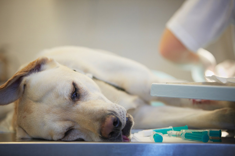
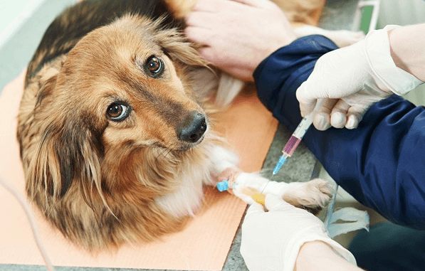
Dirofilariasis requires long-term and expensive treatment, and its effectiveness will depend on the degree of development of the pathology and the location of the parasites in the animal's body. In the subcutaneous form, surgical intervention is necessary to remove adult helminths. After this, the internal and external surfaces of the wound are treated with a 10% imidacloprid solution or a 2.5% moxidectin solution. This procedure is usually sufficient to completely eliminate the parasites. Some specialists recommend treating subcutaneous dirofilariasis with Fenisthion drops applied to the withers at a rate of 20 mg/kg of body weight (for the first three days). After this, a month-long break is required, and treatment is continued for another four days.
The cardiac form of the disease is much more difficult to treat, and treatment is not always effective. Dirofilariasis can be fatal. While medications such as Ivermectin kill worm larvae, they are ineffective against adult worms. Using such medications in the presence of adult worms is very dangerous. A dog's weakened body may not be able to withstand the toxins released by the dead worms. Instead of the expected relief, the condition may only worsen. In particularly severe cases, surgical removal of the parasites from the heart is recommended.
The operation is performed and worms are removed in two ways:
- Through the vena cava of the atrium.
- Using special forceps and X-ray and ultrasound.
Even if the animal has no history of health problems, changes in the dog's condition must be monitored very carefully during treatment with this drug.
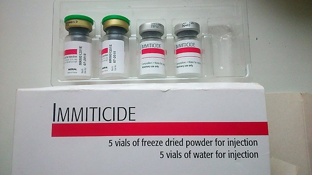
Recently, the intravenous injection solution Thiacetarsamide has been widely used in the fight against dirofilariasis. The treatment course is approximately two weeks, administered twice daily. However, there are some caveats, as the drug only affects adult parasites and does not kill the larvae. Therefore, the treatment is repeated after a certain period of time. Immiticide, a toxic arsenic-based agent, has a similar effect. Both medications are equally dangerous and are not prescribed for treating dogs with liver, heart, kidney, or lung damage. The death of the parasites can lead to pulmonary artery occlusion and severe liver damage. If contraindicated, treatment is carried out with Ivermectin.
For the prevention and treatment of dirofilariasis, your veterinarian may also prescribe the new combination drug Dironet (based on ivermectin, pyrantel pamoate, and praziquantel). The drug is available in tablets and suspension.
Possible complications and prognosis
When subcutaneous dirofilariasis is diagnosed, the prognosis is favorable. The main thing is to identify the pathology in a timely manner and begin treatment as quickly as possible. It's much more dangerous when parasites lodge in the animal's heart. After all, both the medications used and the toxins released by the dead worms are equally detrimental to the dog's body. Even if the animal is saved, it will require lengthy and arduous rehabilitation.
Prevention
It's impossible to completely protect your pet from infection, but you can minimize the risks. Before going outside during warmer months, treat your dog with repellent sprays. Avoid taking your dog for walks during periods of high mosquito populations, and install mosquito nets on your windows. Regularly (every 1-2 months) apply special anti-parasite drops to the withers, such as Advocate or IN-AP Complex.

Dogs should be treated with filariacidal drugs 2-5 months before the mosquito season, and then during the insect season, treatments should be carried out for 5-7 days every 45 days.
Some veterinarians suggest vaccinating with Diethylcarbamazine and giving the animal anthelmintic drugs for preventative purposes.
Dirofilariasis in dogs on video
Reviews
There are good preventative measures for this disease. Those same drops, sprays, and other medications from the veterinary pharmacy labeled as mosquito repellents! This disease is transmitted exclusively by mosquito bites. Prevention is better than cure. And it can be cured—for example, a friend of mine brought a German back from the brink of death. Be sure to treat your animals during periods when mosquitoes, ticks, and other pests are active.
I know that medications are really toxic. Treatment carries the risk of embolism (mechanical blockage of a cardiac or pulmonary artery by a dead bug). This can happen without treatment, but it can also happen if the bug dies of old age in an inappropriate place. That's if it's cardiopulmonary. If it's subcutaneous, well, it encapsulates and that's it. I currently have an elderly woman with dirofilariasis, Aza. So, we've given it a miss. It's not the biggest problem. I'm afraid to think how old my grandmother is, and I don't want to risk liver failure. Any decision should be made carefully, based on the patient's characteristics. IMHO. And dirofilariasis seems to be becoming a very fashionable disease.
This dangerous disease, although difficult to treat, is treatable. The earlier it is detected, the greater the chance of a full recovery. To avoid problems, it's important to take preventative measures seriously. After all, any disease is easier to prevent than to cure.